Diffuse astrocytoma
Diffuse astrocytomas are low-grade astrocytomas classified as WHO grade II tumors of the brain. As the name implies, in vivo there is no clear identifiable border between the tumor and surrounding brain tissue. Diffuse astrocytomas are divided into two molecular groups according to IDH status, IDH mutant and IDH wild-type.
Epidemiology
Diffuse astrocytomas are diagnosed with a biphasic age distribution, being commonly found in young adults between the ages of 20-45 (mean 35 years) and a peak in childhood (6-12 years). Tumors diagnosed in children are usually diffuse brainstem gliomas.
MRI features
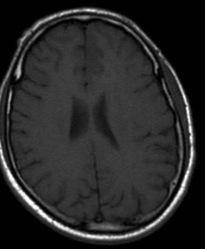
T1: isointense to hypointense compared to white matter, usually confined to the white matter and causes expansion of the adjacent cortex.
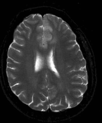
T2/FLAIR: T2 shows a mass-like hyperintense signal
DWI/ADC: lesions typically show facilitated diffusion, with lower ADC values suggesting a higher grade.
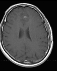
T1 C+ (Gd): no enhancement is often the rule but small ill-defined areas of enhancement are not rare. When enhancement is seen it should be considered as a warning sign for progression to a higher grade tumor.
MR perfusion: There is no elevation of relative cerebral blood volume


